Teomics’s range of ready-to-use histological staining kits includes frequently used staining methods such as Hematoxylin-Eosin, PAS, Elastica van Gieson and Masson-Goldner Trichrome. Staining kits are also provided for special staining techniques like Warthin-Starry, GMS stain, and Congo red stain.
All of our staining kits are manufactured using standard quality control practices, ensuring excellent batch-to-batch consistency. Our staining kits deliver reliable, reproducible results.
$ 99.26
Description
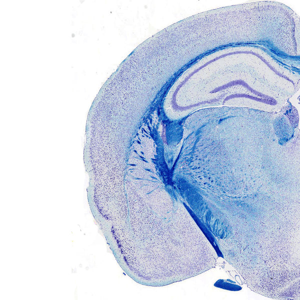
Our kit is designed for staining myelin/myelinated axons and Nissil substance on formalin fixed, paraffin-embedded tissue as well as frozen tissue. Our kit is used for identifying the basic neuronal structure in brain or spinal cord sections, and contains a Cresyl Echt Violet counterstain. Using our protocol and the modified formulation, myelinated tissue can be stained in as little as 30 minutes.
$ 79.92 $ 149.00
Description
Our Hematoxylin and Eosin Staining Kit makes it easy to produce reproducible, high quality H&E histology. The kit contains everything needed for Hematoxylin and Eosin staining and is intended for use in histology and cytology applications. Included in this kit is an optimized formulation of Eosin Y that provides the benefits of a traditional alcoholic formula with significant improvements in usability. Advantages of our kit include lower evaporation rates, better color patterns, a reduced tendency to spill over containers, hands, and countertops, and improved surface tension to remain on tissue section. Mayer's Hematoxylin (Lillie's Modification) produces crisp, intense blue nuclei that provides optimal contrast to the Eosin stained cytoplasm.
Comes with 500 ml bottles (each) of hematoxylin, eosin, and bluing reagent solutions, sufficient for hundreds of slides.
$ 170.00
The Calcium Stain Kit (Modified Von Kossa) is intended for use in the histological visualization of calcium deposits in paraffin or frozen sections. The Modified Von Kossa method is a precipitation reaction in which silver ions react with phosphate (not calcium) in the presence of acidic material. Photochemical degradation of silver phosphate to silver then occurs under UV light illumination. Tissues are treated with a silver nitrate solution and silver replaces calcium reduced by strong light, and so can be visualized as metallic silver. To confirm the presence of calcium, Nuclear Fast Red solution is used to confirm the presence of calcium deposits.
Calcium in mass deposits: Black
Calcium in dispersed deposits: Gray
Nuclei: Red
Cytoplasm: Light Pink
Instructions For Use
SDS
$ 99.00
The Amyloid Stain Kit (Congo Red) is intended for use in the histological visualization of amyloid in tissue sections. Examination under a polarizing microscope results in green birefringence of amyloid.
Amyloid: Red to Pink
Erythrocytes: Light Orange
Eosinophil Granules: Orange to Red
Nuclei: Blue
$ 99.00
The Alcian Blue - PAS Stain Kit is intended for use in the simultaneous histological visualization of sulfated and carboxylated acid mucopolysaccharides, sulfated and carboxylated sialomucins (glycoproteins) and neutral mucins.
Acidic Sulfated Mucosubstances: Blue
Hyaluronic Acid: Blue
Sialomucins: Blue
Neutral Mucins: Magenta
Mixtures of Acidic and Neutral Mucins: Blue - Mauve (depending on the dominant entity)
$ 49.00
The Papanicolaou (PAP) Stain Kit is designed to differentiate between a variety of cells in vaginal smears for detection of vaginal, uterine and cervical cancer. In addition, this procedure is valuable for staining a variety of other bodily secretions and cell smears. The procedure was developed in the early 1940's by George Papanicolaou.
Nuclei: Blue
High Keratin Cells: Orange
Superficial Cells: Pink
Erythrocytes: Dark
Pink Parabasal Cells: Blue/Green
Intermediate Cells: Blue/Green
Metaplastic Cells: May contain both Blue/Green and Pink.
$ 173.00
Mucicarmine Stain Kit (Modified Southgate's) is intended for use in the histological visualization of acid mucopolysacharides in tissue sections. This product is useful in distinguishing mucin negative undifferentiated squamous cell lesions from mucin positive adenocarcinomas. In addition, this product will stain the mucopolysaccharide capsule of Cryptococcus neoformans.
Mucin: Pink/Red
Capsule of Cryptococcus: Red
Nuclei: Black/Blue
Other tissue components: Yellow
$ 499.00
The Jones Stain Kit is intended for use in histological demonstration of the basement membrane and reticular fibers. This procedure is ideal for staining renal glomerular basement membranes. The main function of the basement membrane and reticular fibers is to provide anchorage and support. They are normally found throughout the body, particularly in the kidney, spleen, and lung.
Basement Membrane: Black
Reticulum Fibers: Black
Nuclei: Red
Cytoplasm: Light Pink
$ 32.20
The Gram Stain Kit is intended for the demonstration and differentiation of Gram-positive and Gram-negative bacteria. Gram staining is a common technique used to differentiate between two large groups of bacteria based upon cellular wall composition, coloring Gram-positive and Gram-negative groups red or violet. Gram-positive bacteria will stain violet by retaining crystal violet in a thick layer of peptidoglycan in their cell walls. Gram-negative bacteria will stain red because crystal violet in not retained in the thinner peptidoglycan wall during the decoloring process.
Gram Positive Bacteria: Blue
Gram Negative Bacteria: Red
Other Tissue: Yellow
Nuclei: Red
$ 416.00
The Modified Gomori Methenamine-Silver Nitrate Stain (GMS Stain Kit) is intended for use in the histologic visualization of fungi, basement membrane and some opportunistic organisms such as Pneumocystis carinii. Pneumocystis carinii is an opportunistic pathogen that causes severe pulmonary disease in humans, dogs, rats, mice and other vertebrate species with acquired, induced, or inherited immune deficiency syndromes. In addition, this procedure will demonstrate Actinomyces and related species, Nocardia asteroids, and certain encapsulated bacteria.
$ 185.00
The Fontana-Masson Stain Kit is intended for use in the histological visualization of Argentaffin cells and Melanin in paraffin or frozen sections.
Argentaffin Cells: Black
Melanin: Black
Nuclei: Red
Cytoplasm: Light Pink
$ 215.00
The Eosinophil - Mast Cell Stain Kit is intended for simultaneous visualization of eosinophils and mast cells.
Mast Cells: Bright Blue
Eosinophils: Bright Red
Nuclei: Blue
Control Tissue: Any well-fixed section (3-5 microns) or any air dried smear on a clean slide.
$ 250.00
The Elastic Stain Kit is intended for use in the histological identification of elastin in tissue sections. Demonstration of elastic tissue is useful in cases of emphysema (atrophy of elastic tissue), arteriosclerosis (thinning and loss of elastic fibers) and various other vascular diseases.
Elastic fibers: Black to Blue/Black
Nuclei: Blue to Black
Collagen: Red
Muscle & Other: Yellow
Control Tissue: Lung or any vascular tissue.
$ 215.00
The Copper Stain Kit (For Microwave) is a rapid (2 hr) staining kit that is intended for the demonstration of copper deposits in tissue sections, most commonly in cases of suspected Wilson's disease. Wilson's disease is a rare autosomal disorder caused by decreased serum ceruloplasmin, a blood protein that transports serum copper. Ceruloplasmin deficiency leads to excessive copper accumulation in brain, eye, and liver. In the liver, hepatic copper accumulation results in fatty change, acute hepatitis, chronic hepatitis, eventually resulting in cirrhosis. Urinary copper excretion is increased.
The rubeanic acid and rhodanine stains in the Copper Stain Kit are used to stain the cytoplasmic accumulation of copper in the liver.
Copper Deposits: Light Brown to Red
Nuclei: Blue
$ 164.00
The Colloidal Iron Stain Kit is used for the histological visualization of carboxylated and sulfated mucopolysaccharides and glycoproteins At low pH, colloidal ferric ions are absorbed principally by carboxylated and sulfated mucosubstances. The Prussian blue reaction is used to detect iron bound to the tissue; the stain is differentiated by washing out the excess reagent.
Acid Mucopolysaccharides: Bright Blue
Collagen: Shades of Red
$ 449.00
If you are interested in getting the best quality stain, our Bielschowsky's Stain Kit (Modified) provides for the crisp histological visualization of nerve fibers, neurofibrillary tangles and senile plaques in Alzheimer's disease and other neurodegenerative disorders. This kit has been optimized to allow for variable slide batch sizes, and the developing step can be controlled on a slide-by-slide basis to achieve optimal staining and differentiation. This kit delivers excellent stable staining quality, with minimal overstains, and a low level of background.
This economical kit contains sufficient reagents to stain up to 300 slides.
Axons: Black
Neurofibrillary Tangles: Black
Senile Plaques: Black
Nuclei: Dark Brown
Background: Yellow to Light Brown
$ 75.00
The Alcian Blue (pH 2.5) Stain Kit is intended for use in the histological visualization of sulfated and carboxylated acid mucopolysaccharides and sulfated and carboxylated sialomucins (glycoproteins). Kit contents contain Alcian Blue Solution (pH 2.5), 250ml Acetic Acid Solution, 500ml, and 250 ml of Safranin O Solution.
Acidic Sulfated Mucosubstances: Blue
Hyaluronic Acid: Blue Sialomucins: Blue
Nuclei: Red
Background: Pink
$ 75.00
The Alcian Blue (pH 1.0) Stain Kit is intended for use in the histological visualization of strongly sulfated mucosubstances.
Strongly Sulfated Mucosubstances: Blue
Nuclei: Red
Background: Pink
$ 175.00
The Alcian Blue (pH 0.5) Stain Kit is intended for use in the histological visualization of very strongly sulfated mucosubstances.
Very Strongly Sulfated Mucosubstances: Blue
Nuclei: Red
Background: Pink
$ 119.00
The Acid Fast Bacteria (AFB) Stain Kit is intended for use in the histological visualization of Acid Fast Bacteria and Tubercle Bacilli. This kit is a rapid 15-minute procedure. The lipoid capsule of the acid-fast organism takes up carbol fuchsin and resists decolorization.
Acid Fast Organisms: Bright Red
Background: Light Green